FFPE Tissue Samples and Digital Pathology

Modern biomedical research increasingly depends on access to high-quality biological specimens and detailed pathology information. Among the most valuable resources available to researchers are FFPE tissue samples and H&E scanned slides. These resources provide important information about tissue morphology, disease progression, cancer development, and cellular characteristics.
Formalin-Fixed Paraffin-Embedded, commonly known as FFPE, is one of the most widely used methods for preserving tissue samples. For decades, hospitals, pathology laboratories, research institutions, and biobanks have stored tissues using this method. These preserved specimens can provide researchers with access to valuable historical and clinical material.
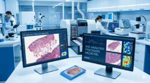
At the same time, advances in digital pathology have transformed traditional glass slides into high-resolution digital images. H&E stained tissue sections can now be scanned and stored in secure digital systems, allowing researchers and pathologists to access tissue images remotely.
A well-organized digital pathology repository can bring together biological specimens, pathology images, clinical information, and research data in a structured environment. This creates significant opportunities for cancer research, drug development, artificial intelligence, biomarker discovery, and precision medicine.
FFPE Tissue Repository: A Valuable Resource for Biomedical Research
An FFPE tissue repository is a structured collection of formalin-fixed, paraffin-embedded tissue specimens. These repositories may contain samples collected from surgical procedures, biopsies, clinical trials, research studies, and diagnostic pathology workflows.
The major advantage of FFPE preservation is its ability to maintain tissue morphology for long periods. As a result, tissue collections created years ago can still provide valuable information for current scientific research.
Researchers can use FFPE specimens for multiple applications, including:
- Histopathological analysis
- Cancer research
- Biomarker identification
- Molecular testing
- Drug development
- Retrospective clinical studies
- Artificial intelligence training
- Translational medicine
A professionally managed FFPE tissue repository helps researchers locate appropriate samples based on disease type, tissue type, patient demographics, treatment history, or other available research criteria.
The availability of organized FFPE collections is particularly important for retrospective research. Scientists may study archived tissue samples to identify patterns that were not previously recognized when the original diagnosis was made.
H&E Scanned Slides for Digital Pathology and Research
H&E scanned slides are digital versions of tissue sections stained using Hematoxylin and Eosin. H&E staining is one of the most commonly used techniques in pathology because it provides clear visual differentiation between cellular structures and tissue components.
Traditionally, pathologists examine H&E-stained tissue sections using optical microscopes. With modern scanning technology, these physical slides can be converted into high-resolution digital images.
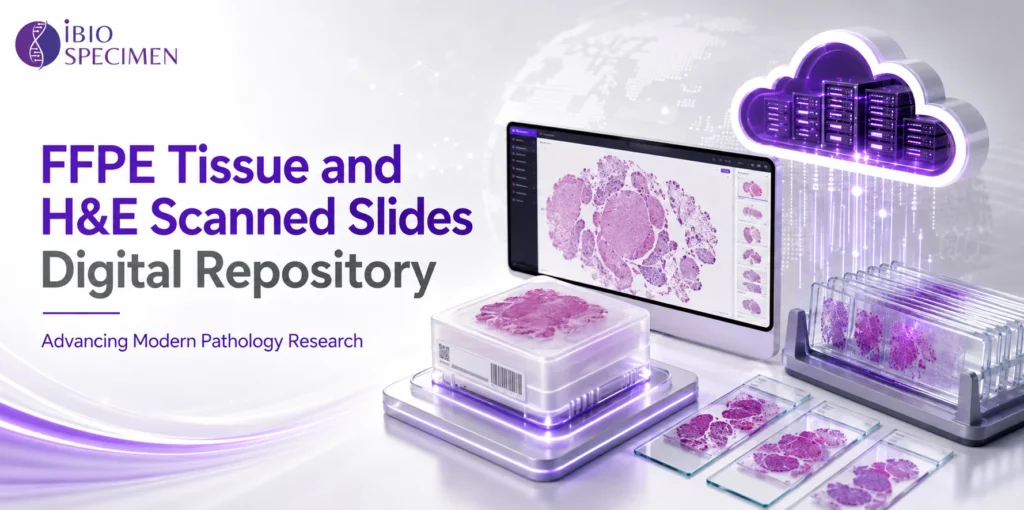
The process of creating H&E scanned slides generally involves preparing the tissue section, staining it with Hematoxylin and Eosin, placing the slide into a high-resolution scanner, and generating a digital whole-slide image.
Digital versions of pathology slides offer several advantages:
- Remote access to pathology images
- Easier collaboration between researchers
- Improved image sharing
- Support for artificial intelligence analysis
- Long-term digital preservation
- Reduced dependence on physical slide transportation
- Faster access to large image collections
H&E scanned slides can also support educational programs. Medical students, pathology trainees, and researchers can study digitized tissue images without requiring physical access to an extensive slide collection.
Digital Pathology Repository for Centralized Research Access
A digital pathology repository provides an organized system for storing, managing, and accessing pathology images and related information. Instead of maintaining isolated collections of physical slides, institutions can create centralized digital environments.
A modern repository may contain:
- Whole-slide images
- H&E scanned slides
- Digital histology slides
- Tissue metadata
- Diagnostic information
- Research annotations
- Imaging data
- Sample identifiers
- Associated molecular information
Centralized storage makes it easier for authorized researchers to search large collections and identify relevant pathology materials.
A digital pathology repository can also support collaboration between institutions. Researchers located in different geographic regions may be able to review the same pathology images without shipping fragile glass slides.
This capability is increasingly valuable for multicenter research studies and international collaborations.
FFPE Tissue Archive and Long-Term Specimen Preservation
An FFPE tissue archive contains preserved tissue specimens that have been collected and stored over time. These archives can represent an important historical record of disease and clinical pathology.
For cancer research, archived tissue may provide access to specimens collected before modern molecular technologies were available. Researchers can revisit these samples using new scientific methods and analytical tools.
For example, a tissue specimen collected years ago may be evaluated today using advanced genomic, proteomic, or image analysis technologies.
An FFPE tissue archive can therefore support:
- Retrospective cancer studies
- Longitudinal disease research
- Biomarker validation
- Historical pathology analysis
- Drug response studies
- Rare disease investigations
Proper organization and documentation are essential for maximizing the scientific value of archived tissue collections.
Histopathology Slide Database for Efficient Information Management
A histopathology slide database allows pathology images and associated information to be organized in a searchable system.
Large pathology departments and research institutions may generate thousands of slides over time. Without a structured database, locating specific images or specimens can become difficult.
A well-designed histopathology slide database may allow users to search based on criteria such as:
- Disease type
- Tissue type
- Cancer subtype
- Staining method
- Research category
- Sample availability
- Image characteristics
- Collection date
The ability to search and filter information can significantly improve research efficiency.
Instead of manually reviewing large numbers of physical slides, researchers can identify potentially relevant materials through a structured digital system.
Whole Slide Imaging Repository for High-Resolution Analysis
A whole slide imaging repository stores complete digital images of pathology slides.
Whole-slide imaging technology captures an entire tissue section at high resolution. Unlike traditional photographs that show only selected areas, whole-slide images allow users to navigate across the entire tissue specimen digitally.
Researchers can zoom into specific cellular structures and examine different regions of the tissue.
A whole slide imaging repository can support:
- Digital pathology review
- AI model development
- Quantitative image analysis
- Remote pathology consultation
- Educational programs
- Cancer research
- Collaborative studies
The growing adoption of whole-slide imaging is contributing to the transformation of pathology from a primarily microscope-based discipline into a data-rich digital environment.
Digitized H&E Slides for Remote Collaboration
Digitized H&E slides provide researchers and pathologists with flexible access to tissue images.
In traditional pathology workflows, physical slides often need to be transported between laboratories or institutions. This can create logistical challenges and increase the risk of damage or loss.
Digitized slides reduce many of these limitations.
Authorized users can review images through secure digital systems, collaborate with colleagues, and share research findings more efficiently.
Digitized H&E slides are particularly useful for international research collaborations. Multiple experts can review the same tissue image and provide independent interpretations.
This can support:
- Expert consultation
- Research collaboration
- Diagnostic quality review
- Educational training
- AI-assisted image analysis
Digital access also creates opportunities for more standardized pathology workflows.
Pathology Tissue Samples Supporting Translational Research
Pathology tissue samples connect laboratory research with real-world disease.
These samples can provide direct information about cellular morphology, tissue organization, tumor characteristics, inflammation, and disease progression.
Researchers often use pathology tissue samples to investigate questions such as:
- How does a disease develop?
- Which biomarkers are associated with disease progression?
- How do tumors respond to treatment?
- What cellular characteristics predict patient outcomes?
- Which patients may benefit from specific therapies?
When tissue samples are linked with appropriate clinical and research information, their scientific value can increase significantly.
However, responsible data management and ethical governance are essential when handling human biological materials.
FFPE Specimen Database for Organized Sample Discovery
An FFPE specimen database helps researchers identify and manage preserved tissue materials.
The database may contain information about the tissue source, disease category, preservation method, specimen characteristics, and available supporting data.
A structured FFPE specimen database can improve sample discovery and reduce the time required to locate appropriate research materials.
For research organizations, effective sample management can help prevent duplication and improve the utilization of valuable biological resources.
An advanced database may also integrate digital pathology images with specimen information.
This creates a stronger connection between the physical tissue sample and its corresponding visual pathology data.
Digital Histology Slides Transforming Pathology Workflows
Digital histology slides are becoming increasingly important in research, education, and diagnostic pathology.
Digital systems allow pathology images to be viewed on computer workstations and specialized software platforms.
These technologies can support advanced image analysis techniques, including automated cell detection, tissue segmentation, pattern recognition, and quantitative measurement.
Digital histology slides are also supporting the development of artificial intelligence applications.
Machine learning systems can analyze large collections of annotated pathology images to identify patterns associated with disease.
Potential applications include:
- Tumor detection
- Cell classification
- Tissue segmentation
- Biomarker analysis
- Disease prediction
- Research automation
The availability of high-quality, well-organized digital histology datasets is essential for developing reliable AI systems.
Cancer Tissue Repository for Oncology Research
A cancer tissue repository can provide researchers with access to tissue specimens representing different tumor types and disease stages.
Cancer is highly complex and biologically diverse. Even tumors classified under the same diagnosis may demonstrate significant differences at the molecular and cellular levels.
Access to diverse cancer tissue collections allows researchers to study this variation.
A cancer tissue repository may support research involving:
- Breast cancer
- Lung cancer
- Colorectal cancer
- Prostate cancer
- Ovarian cancer
- Brain tumors
- Rare cancers
Cancer tissue resources can contribute to biomarker research, drug development, treatment response studies, and precision oncology.
When combined with digital pathology images, cancer tissue repositories can provide a powerful research environment.
Researchers can analyze both the biological specimen and its visual tissue characteristics.
Pathology Image Database for AI and Image Analysis
A pathology image database is increasingly valuable for computational pathology.
Artificial intelligence systems require large and diverse datasets for training and validation.
Pathology images can be used to train algorithms to recognize cellular structures, identify abnormal tissue patterns, and support quantitative analysis.
However, the quality of the dataset is extremely important.
A useful pathology image database should include well-organized images, appropriate metadata, consistent image quality, and reliable annotation where applicable.
Diverse datasets are also important for improving the generalizability of AI models.
A system trained using images from only one laboratory or scanner may not perform equally well across other clinical environments.
For this reason, digital pathology repositories with diverse image collections can play an important role in the future development of AI-powered pathology tools.
H&E Slide Scanning and the Digital Transformation of Pathology
H&E slide scanning is the process of converting traditional glass pathology slides into high-resolution digital images.
Modern scanners capture tissue sections at different magnification levels and generate files that can be viewed using specialized software.
The digital transformation created by H&E slide scanning offers significant benefits.
These include improved access, easier collaboration, reduced physical handling, and support for computational analysis.
For research institutions, scanning existing pathology collections can help preserve valuable information and create new opportunities for data-driven research.
However, successful digitization requires careful quality management.
Image quality, focus, staining consistency, scanning resolution, and metadata accuracy can all affect the usefulness of digital slides.
Digital Tissue Specimen Repository for the Future of Research
A digital tissue specimen repository represents the combination of biological specimen management and digital information systems.
Rather than treating tissue samples and pathology images as separate resources, modern repositories can connect physical specimens with digital images and research data.
This integrated approach can improve:
- Sample traceability
- Research collaboration
- Data accessibility
- AI development
- Biomarker discovery
- Study reproducibility
A digital tissue specimen repository may become an important part of future biomedical infrastructure.
As imaging technologies, artificial intelligence, and molecular analysis continue to advance, researchers will increasingly require integrated systems capable of managing complex biological datasets.
The Role of Digital Repositories in Artificial Intelligence
Artificial intelligence is one of the most significant emerging applications for digital pathology.
AI systems can process large numbers of whole-slide images and identify patterns that may be difficult or time-consuming to evaluate manually.
However, AI development depends on access to high-quality datasets.
Repositories containing FFPE tissue samples, digitized H&E slides, and associated pathology information can help support the development of machine learning models.
AI applications may assist researchers with:
- Image classification
- Tissue segmentation
- Cell counting
- Tumor identification
- Pattern recognition
- Quantitative pathology
Importantly, AI should be developed and validated carefully before being used in clinical decision-making.
High-quality repositories can support this process by providing structured datasets for research and validation.
Benefits of Combining Physical and Digital Pathology Resources
The greatest research value may come from combining physical specimens with digital information.
For example, an FFPE tissue sample may be connected with:
- H&E scanned slides
- Whole-slide images
- Histopathology information
- Molecular data
- Research annotations
This creates a more comprehensive research resource.
Researchers can initially identify interesting patterns within digital pathology images and then investigate corresponding biological material.
This workflow can support biomarker discovery and translational research.
The integration of physical and digital resources also makes it easier to manage complex research programs involving multiple laboratories and institutions.
The Future of FFPE Tissue and Digital Pathology Repositories
The combination of FFPE tissue samples, H&E scanned slides, and advanced digital systems is transforming biomedical research and pathology.
An organized FFPE tissue repository can preserve valuable biological materials, while a digital pathology repository can make pathology information more accessible and useful.
Technologies such as H&E slide scanning, whole-slide imaging, and digital image analysis are creating new opportunities for cancer research, artificial intelligence, and precision medicine.
A well-managed FFPE tissue archive, histopathology slide database, whole slide imaging repository, and digital tissue specimen repository can help researchers access information more efficiently and collaborate across geographic boundaries.
As biomedical research continues to generate larger and more complex datasets, digital repositories will become increasingly important.
The integration of digitized H&E slides, pathology tissue samples, FFPE specimen databases, digital histology slides, cancer tissue repositories, and pathology image databases represents an important step toward a more connected and data-driven future for pathology research.
Organizations that invest in secure, well-organized, and scientifically valuable digital pathology infrastructure will be better positioned to support the next generation of research, artificial intelligence, and precision healthcare.