
In the traditional drug discovery workflow, the gap between the physical tissue specimen and the data scientist has been a significant bottleneck. Researchers would procure FFPE (Formalin-Fixed Paraffin-Embedded) tissue blocks, wait for international shipping, cut slides in their own labs, and then digitize them for analysis.
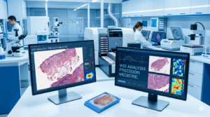
At iBiospecimen.com, we have eliminated this friction. By providing Human Tumor Blocks with High-Resolution Digital Pathology Slides (Whole Slide Images – WSI) included, we offer a “Digital-First” solution for the modern biotech era. This integrated approach allows research teams to begin their computational analysis, AI model training, and pathologist reviews the moment they place an order—long before the physical block arrives at their facility.
- What is a “Digital-Ready” Human Tumour Block?
A “Digital-Ready” biospecimen is more than just a piece of tissue. It is a dual-asset procurement model consisting of:
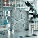
- The Physical FFPE Block:A high-quality, pathologically verified tumor specimen processed under strict pre-analytical standards (Cold Ischemia Time $<60$ minutes).
- The Whole Slide Image (WSI):A diagnostic-grade, high-resolution scan (typically 20x or 40x magnification) of a representative H&E-stained section from that specific block.
- Integrated Metadata:De-identified clinical history, mutation reports (NGS), and IHC status, all linked via a secure digital ID.
- The Strategic Advantage for Biotech and AI Foundries
For companies building AI foundries or developing deep-learning algorithms for clinical diagnostics, the inclusion of digital pathology slides is a force multiplier.
- Training “Ground Truth” AI Models
AI is only as good as the data it consumes. Our digital slides provide the “Ground Truth” needed to train machine learning models to:
- Identify Morphological Patterns:Recognize rare tumor subtypes or features like Perineural Invasion (PNI) and Lymphovascular Space Invasion (LVSI).
- Automate Grading:Develop algorithms for Gleason scoring in prostate cancer or Nottingham grading in breast cancer.
- Predict Genotypes:Train models to identify specific genetic mutations (e.g., EGFR or TP53) directly from the H&E slide morphology.
- Remote Pathologist Review and Collaboration
With digital slides, your primary investigators and consulting pathologists can review the tissue from anywhere in the world. This facilitates:
- Pre-Selection:Review the digital slide before the block is shipped to ensure the tumor content and necrosis levels meet your study’s strict inclusion criteria.
- Inter-Observer Consensus:Allow multiple pathologists to score the same digital slide simultaneously to ensure reproducibility in your study data.
- Technical Specifications: Scan Quality and Compatibility
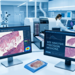
At iBiospecimen.com, we utilize state-of-the-art scanning technology to ensure our digital assets meet the rigorous demands of computational pathology.
Resolution and Magnification
- 20x and 40x Scans:We offer scanning at $2 (20x) or $2.5 (40x), providing the clarity needed to see nuclear detail and mitotic figures.
- Z-Stacking:For samples with significant architectural complexity, we offer multi-plane Z-stack scanning to ensure focus throughout the tissue depth.
File Formats and Interoperability
Our digital pathology slides are provided in open-standard and vendor-neutral formats to ensure they integrate seamlessly with your existing software:
- .SVS (Aperio)
- .NDPI (Hamamatsu)
- .TIFF / BigTIFF
- DICOM Compliance:We are transitioning toward full DICOM compatibility to support the next generation of clinical imaging standards.
- Featured Inventory: AI-Ready Tumor Cohorts
We maintain a worldwide repository of human tumor blocks across all major oncology indications, each paired with its digital counterpart.
| Indication | Digital Feature Focus | Metadata Included |
| Non-Small Cell Lung Cancer | Tumor Microenvironment (TME) mapping | EGFR/ALK/KRAS Status |
| Breast Carcinoma | TILs (Tumor Infiltrating Lymphocytes) | ER/PR/HER2 & Ki-67 |
| Colorectal Cancer | Budding and invasive front analysis | MSI/MMR & BRAF |
| Prostate Adenocarcinoma | Glandular architecture and Gleason grading | PSA & Stage |
| Rare Sarcomas | Mesenchymal cellularity and fusions | Molecular Translocation data |
- Bridging the Gap: From Digital Scans to Spatial Biology
While digital pathology provides the morphological landscape, the physical block remains essential for “Spatial Multi-Omics.”
Spatial Transcriptomics Integration
Our digital slides serve as the “scout image” for spatial transcriptomics platforms like 10x Genomics Visium or Nanostring GeoMx. Researchers can use the digital slide to identify specific “Regions of Interest” (ROIs)—such as the leading edge of a tumor or an immune-rich niche—and then target those exact coordinates on the physical block for molecular analysis.
Digital-to-Molecular Correlation
By pairing a digital pathology slide with an NGS mutation report, researchers can correlate visual phenotype (the “Pathomics”) with the genetic genotype (the “Genomics”). This dual-layered data is the foundation of modern precision medicine.
- Pre-Analytical Excellence: Protecting the Image Quality
A digital scan is only as good as the tissue it represents. Our blocks are processed under a strict “Pathology Excellence” protocol:
- Optimal Fixation:10% Neutral Buffered Formalin (NBF) to prevent “formalin pigment” or “acid hematin” artifacts that can interfere with AI color normalization.
- Precision Sectioning:We utilize microtomes calibrated to 3–5 $\mu$m thickness to ensure a flat, uniform section that remains in focus across the entire scan area.
- Clean Slide Preparation:We ensure slides are free of air bubbles, dust, or mounting medium artifacts that can create “blind spots” for AI algorithms.
- Ethical Standards and Global Digital Logistics
The transfer of digital pathology data is subject to the same rigorous ethical standards as physical tissue.
- Anonymization:All digital slides are de-identified. Patient names and hospital IDs are removed from the digital labels and replaced with a unique iBiospecimen GUID (Globally Unique Identifier).
- Cloud-Based Delivery:We offer secure, encrypted cloud delivery of WSIs via high-speed AWS or Google Cloud gateways, allowing for instant access upon purchase.
- Regulatory Compliance:Fully compliant with HIPAA and GDPR for the storage and transmission of medical imaging data.
- Why iBiospecimen.com is Your Strategic Partner in Digital Pathology
We understand that you aren’t just buying tissue; you are buying the ability to generate insights.
- “Pre-Scan” Inventory Access:Browse our digital library before you buy. See the morphology and tumor content of the block before committing your budget.
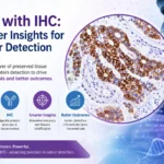
- Custom Scanning Services:Need a specific stain? We can perform IHC (e.g., PD-L1, HER2) on your selected block, scan the result, and provide the IHC-WSI paired with the H&E-WSI.
- Data Consistency:Every block and slide is accompanied by a Certificate of Analysis (CoA), ensuring the technical specifications of the scan match your study requirements.
Conclusion: The Era of the Digital Biobank
The integration of human tumor blocks with digital pathology slides represents a paradigm shift in biospecimen procurement. It empowers the researcher with the speed of digital data and the depth of physical biology. At iBiospecimen.com, we are proud to be the bridge between the clinic and the computer, providing the high-fidelity material your AI models and molecular assays require.
Frequently Asked Questions (FAQs)
Q: Can I download the digital slides before the physical blocks ship?
A: Yes. Once your purchase is confirmed, we provide a secure link to download the Whole Slide Images (WSI) in your preferred format (.svs, .ndpi, etc.).
Q: Are your digital slides compatible with AI software like QuPath or Visiopharm?
A: Absolutely. Our vendor-neutral formats are designed to be imported directly into all major open-source and commercial digital pathology analysis platforms.
Q: What is the typical tumor purity of your digital-ready blocks?
A: We prioritize blocks with >30% tumor nuclei. The exact tumor percentage is assessed by a pathologist on the digital slide and provided in the metadata.
Keywords: Human Tumor Blocks, Digital Pathology Slides, Whole Slide Imaging (WSI), AI-Ready Biospecimens, Digital Biobank, Oncology Tissue Scans, iBiospecimen, SVS Pathological Slides, Remote Pathology Review.
Unlock the power of digital oncology. Contact the procurement team at iBiospecimen.com today to access our digital pathology library and order your sequenced tumor blocks.