
Whole Slide Scanning Services
Advanced Whole Slide Scanning Services for Researchers and Industry
DIGITAL PATHOLOGY SLIDE SCANNING
At iBioSpecimen, we provide premium whole slide scanning services for pathology laboratories, pharmaceutical companies, CROs, biotech firms, academic institutions, diagnostic companies, and research organizations worldwide. Our advanced digital pathology scanning services help convert traditional glass slides into high-resolution digital pathology images that can be used for pathology review, image analysis, research collaboration, digital archiving, AI model development, telepathology workflows, and clinical research support.
Whole slide imaging is transforming the way pathology slides are reviewed, shared, stored, and analyzed. By digitizing glass slides into clear and detailed digital images, organizations can improve accessibility, reduce dependency on physical slide movement, support remote pathology review, and create structured digital pathology archives. iBioSpecimen helps research and clinical teams unlock greater value from their pathology slide collections by offering reliable, scalable, and secure pathology slide scanning services tailored to each project’s requirements.
What Is Whole Slide Scanning and How Does It Work?
Whole slide scanning is the process of converting traditional glass pathology slides into high-resolution digital images that can be viewed, shared, analyzed, and stored electronically. Instead of reviewing a physical slide only under a microscope, whole slide imaging allows the entire tissue section to be captured as a digital pathology image for remote review, research, teaching, archiving, and advanced image analysis.
The process begins with preparing a stained glass slide, such as an H&E slide, IHC slide, special stain slide, cytology slide, or FFPE tissue section. The slide is then loaded into a whole slide scanner, which captures detailed images of the tissue at high magnification, commonly at 20x or 40x. These images are digitally stitched together to create a complete whole slide image that can be viewed on a computer through digital pathology software.
Once scanned, the digital pathology image can be used by pathologists, researchers, pharmaceutical companies, CROs, biotech firms, academic institutions, and diagnostic developers for multiple applications. These include pathology review, tumor assessment, biomarker analysis, IHC scoring, clinical trial support, telepathology, digital archiving, AI model training, and computational pathology research.
Whole slide scanning helps organizations reduce the need to repeatedly ship fragile glass slides, improves collaboration between remote teams, supports faster slide review, and creates long-term digital pathology archives. For research and pharmaceutical projects, high-quality whole slide images can also support oncology studies, companion diagnostic development, biomarker discovery, and AI-based pathology workflows.
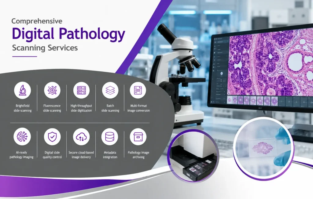
Complete Digital Pathology Imaging and Scanning Services
Our digital pathology scanning services support a broad range of pathology, biospecimen, research, and clinical development applications. We help laboratories, pharmaceutical companies, CROs, biotech firms, academic institutions, and diagnostic organizations convert glass slides into high-quality digital pathology images for review, analysis, archiving, AI development, and collaborative research.
Our services include brightfield slide scanning, fluorescence slide scanning, high-throughput slide digitization, batch slide scanning, multi-format image conversion, AI-ready pathology imaging, digital slide quality control, secure cloud-based image delivery, metadata integration, and pathology image archiving.
iBioSpecimen specializes in accurate, reliable, and scalable pathology image digitization for both small research projects and large enterprise-level slide scanning programs. Whether you need a limited number of slides digitized for a pilot study or thousands of pathology slides scanned for clinical research, AI model training, or digital archiving, our team provides flexible digital pathology imaging solutions tailored to your project requirements.
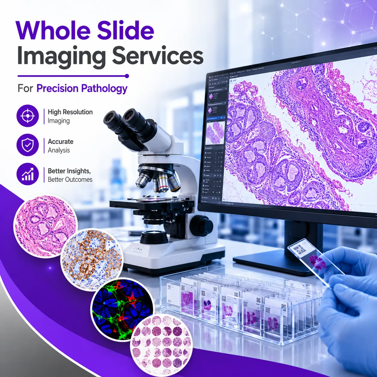
High-Resolution Whole Slide Imaging Services for Precision Pathology
Our whole slide imaging services are designed for research institutions, pathology laboratories, hospitals, pharmaceutical companies, CROs, biotechnology organizations, and diagnostic developers that require reliable, high-quality, and scalable digital pathology workflows. By converting traditional glass slides into high-resolution digital slide images, iBioSpecimen helps organizations improve pathology review, streamline collaboration, support research analysis, and build secure digital pathology archives.
Whole slide imaging allows pathology teams and researchers to access complete digital versions of H&E, IHC, special stain, cytology, and FFPE tissue slides without depending solely on physical microscope review. These digital images can be used for remote pathology consultation, tumor assessment, biomarker evaluation, IHC scoring, tissue morphology review, clinical trial support, and multi-site research collaboration.
At iBioSpecimen, our whole slide imaging workflow is built to support accuracy, image clarity, consistency, and efficient project execution. We assist with slide scanning at required magnifications, digital image generation, image quality checks, file organization, metadata integration, and secure digital delivery. Whether your project involves a small batch of research slides or a large-scale pathology digitization program, our team can provide flexible imaging support based on your technical and study requirements.
Our whole slide imaging services for precision pathology are especially valuable for oncology research, companion diagnostic development, biomarker discovery, pharmaceutical R&D, AI pathology model training, and digital pathology platform development. High-quality whole slide images help researchers evaluate tissue architecture, detect disease patterns, analyze biomarker expression, and generate image datasets suitable for advanced computational pathology and machine learning applications.
We support organizations seeking dependable digital pathology workflows for slide review, digital archiving, telepathology, clinical research, and AI-ready pathology imaging. With a focus on image quality, sample traceability, secure handling, and scalable delivery, iBioSpecimen helps transform glass slides into valuable digital pathology assets that can support research, diagnostics, and innovation in precision pathology.
We provide scanning support for multiple slide and tissue types, including:
- FFPE tissue sections
- Frozen tissue slides
- Cytology slides
- Hematology slides
- Immunohistochemistry (IHC) slides
- Fluorescence slides
- H&E stained slides
- Special stain pathology slides
- Tissue microarrays (TMAs)
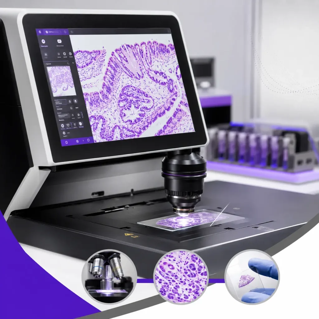
High Resolution Slide Scanning for Accurate Image Capture
Image quality is critical in digital pathology. Our high resolution slide scanning solutions capture fine tissue structures with exceptional clarity and precision.
High-magnification scanning
Accurate tissue reproduction
Sharp cellular detail
Multi-layer focal imaging
Color-calibrated imaging
Large tissue area capture
Consistent image quality control
Our digital pathology workflows are designed to maintain specimen integrity while ensuring reliable image visualization for research and diagnostic applications.
Pathology Slide Scanning Services for Research & Clinical Applications
Our specialized pathology slide scanning services support a broad range of medical and scientific applications.
Our histopathology slide digitization services help laboratories transition from traditional microscopy to scalable digital pathology systems.
Benefits of Histopathology Digitization
- Reduced physical slide handling
- Faster pathology review
- Simplified image sharing
- Enhanced collaboration
- Improved data management
- AI-compatible workflows
- Long-term digital archiving
Modern biospecimen workflows increasingly rely on standardized digital pathology processes to improve research quality and specimen accessibility.
Our secure virtual slide scanning services allow pathologists and researchers to review pathology slides from anywhere in the world.
Digital slides can be accessed through secure cloud platforms, enabling:
- Remote pathology consultations
- Multi-user collaboration
- Real-time image review
- Cross-site pathology discussions
- Education and training
- Digital pathology conferences
Virtual pathology systems reduce operational delays while improving accessibility for global research teams.
We offer scalable tissue slide scanning services for biomedical, translational, and pharmaceutical research applications.
Tissue Types Supported
- Tumor tissues
- Normal tissues
- Rare disease tissues
- Inflammatory tissue samples
- Neurological tissues
- Cardiovascular tissues
- Liver tissues
- Kidney tissues
- Lung tissues
- Gastrointestinal tissues
Our workflows ensure accurate and reproducible medical slide digitization services for research organizations worldwide.
Our glass slide scanning services support both individual projects and high-volume enterprise pathology digitization programs.
Slide Formats Supported
- Standard pathology slides
- Cytology slides
- TMA slides
- Frozen section slides
- Fluorescence slides
- Special stain slides
Each slide undergoes strict quality review to ensure optimal scanning performance and digital image integrity.
Our enterprise-grade digital slide imaging solutions are designed to help laboratories modernize pathology workflows and improve operational efficiency.
Laboratory Benefits
- Streamlined pathology review
- Centralized image management
- Improved research collaboration
- Faster data retrieval
- AI integration capabilities
- Reduced storage burden
- Enhanced pathology accessibility
Digital pathology transformation continues to improve laboratory productivity and pathology data management.
Our pathology image digitization services support next-generation computational pathology initiatives.
Digital pathology datasets can be integrated into:
- AI training workflows
- Machine learning pipelines
- Image segmentation tools
- Biomarker analysis platforms
- Predictive pathology systems
- Quantitative pathology software
We help organizations build scalable pathology imaging datasets for advanced computational research.
Artificial intelligence is transforming pathology. Our AI pathology slide scanning services provide optimized digital pathology images suitable for algorithm development and AI-assisted diagnostics.
AI-Compatible Features
- High-resolution image capture
- Uniform scanning quality
- Consistent color normalization
- Metadata integration
- Large dataset support
- Batch scanning capabilities
AI-ready digital slides improve the efficiency of mac
Our microscopy slide scanning services support a variety of scientific and laboratory imaging applications.
Applications Include
- Cellular imaging
- Histology review
- Immunohistochemistry analysis
- Fluorescence microscopy
- Pathology consultation
- Tissue morphology analysis
We provide accurate digital image capture for both research and diagnostic environments.
Healthcare organizations increasingly rely on medical slide digitization services to improve pathology accessibility and workflow efficiency.
Advantages of Medical Slide Digitization
- Faster pathology turnaround
- Enhanced clinical collaboration
- Improved image accessibility
- Simplified pathology storage
- Reduced risk of slide damage
- Secure digital backups
Digital pathology also supports educational programs and multidisciplinary clinical collaboration.
Our digital pathology workflows support whole slide image analysis for quantitative and computational pathology applications.
Image Analysis Applications
- Cell quantification
- Biomarker scoring
- Tumor segmentation
- Tissue classification
- Morphometric analysis
- AI-assisted diagnostics
Researchers can integrate digital pathology images into advanced image analysis platforms for scalable pathology analytics.
Our diagnostic slide scanning services support pathology labs seeking reliable digital pathology imaging solutions.
Diagnostic Applications
- Histopathology review
- Oncology diagnostics
- Consultation workflows
- Pathology archiving
- Digital case sharing
High-quality digital slides help improve workflow efficiency and facilitate rapid pathology consultations.
We provide scalable research slide scanning services for pharmaceutical, biotech, and academic organizations.
Research Areas Supported
- Cancer research
- Biomarker discovery
- Drug response studies
- Translational medicine
- Rare disease research
- Precision medicine
- Immunology studies
Research institutions benefit from secure digital pathology workflows and centralized slide access systems.
Our advanced histology slide scanning services help researchers digitize tissue sections for image analysis, archival storage, and collaborative review.
Histology Applications
- Tissue morphology evaluation
- Histopathological analysis
- Biomarker validation
- Cellular structure studies
- Tissue architecture analysis
Digital histology improves workflow efficiency while enabling scalable pathology research.
At iBioSpecimen, we support complete pathology digital transformation services for laboratories transitioning to digital pathology infrastructure.
Digital Transformation Capabilities
- Slide digitization programs
- Workflow modernization
- Cloud image management
- Digital pathology integration
- AI pathology enablement
- Data standardization
- Centralized pathology archives
Our solutions help laboratories modernize pathology operations while supporting long-term scalability.
Our outsourced whole slide scanning services help organizations reduce infrastructure costs while accessing enterprise-level pathology digitization capabilities.
Why Outsource Whole Slide Scanning?
- Reduced capital investment
- Faster project turnaround
- Access to expert imaging teams
- Scalable slide processing
- Secure image management
- Flexible project support
Whether you need a few hundred slides or large-scale pathology digitization, iBioSpecimen provides reliable outsourced digital pathology support tailored to your project requirements.
Global Research Support
We support research organizations, CROs, pharmaceutical companies, and academic institutions worldwide.
High-Quality Digital Imaging
Our advanced scanners provide exceptional high resolution slide scanning with reliable image consistency.
Fast Turnaround Times
We deliver efficient pathology digitization workflows for time-sensitive research and diagnostic projects.
Scalable Infrastructure
From pilot studies to enterprise-level pathology programs, our services scale according to project needs.
Secure Data Management
We maintain secure digital pathology workflows with protected image storage and controlled data access.
AI-Ready Pathology Solutions
Our services support next-generation computational pathology and AI-driven image analysis applications.
Need reliable whole slide scanning services for research, diagnostics, AI development, or digital pathology transformation?
iBioSpecimen Whole Slide Services provides advanced digital pathology scanning services, scalable whole slide imaging services, and enterprise-grade pathology digitization solutions for laboratories and research organizations worldwide.
Contact our team today to discuss your project requirements and receive customized digital pathology support.
High-Resolution Digital Pathology Slide Scanning Services for Research, AI Development, and Clinical Workflows
iBioSpecimen provides advanced digital pathology slide scanning services for pharmaceutical companies, biotechnology firms, CROs, pathology laboratories, diagnostic companies, academic institutions, AI companies, and research organizations worldwide. Our whole slide imaging and pathology slide digitization services help convert traditional glass slides into high-resolution digital pathology images that can be used for research analysis, biomarker evaluation, remote pathology review, artificial intelligence development, machine learning model training, clinical trial support, and long-term digital archiving.
Digital pathology is transforming the way tissue slides are reviewed, shared, analyzed, and stored. Instead of relying only on physical glass slides and microscope-based review, organizations can now access complete digital images of H&E, IHC, special stain, cytology, and FFPE tissue slides. These high-quality digital pathology images can be viewed remotely, shared securely with collaborators, integrated into AI workflows, and used to create structured pathology image datasets for advanced research and diagnostic development.
At iBioSpecimen, we support both small-scale and large-scale pathology slide scanning projects, including single-batch research slide scanning, archived FFPE slide digitization, clinical trial pathology scanning, oncology slide scanning, IHC slide scanning, H&E slide scanning, AI-ready whole slide image generation, and enterprise-level digital pathology dataset creation. Our goal is to help organizations accelerate pathology workflows, improve collaboration, reduce physical slide handling, and unlock the full research value of tissue-based biospecimens.
Our whole slide imaging services are designed to generate high-resolution digital images from glass pathology slides using advanced slide scanning technology and structured imaging workflows. Whole slide imaging captures the entire tissue section and converts it into a digital slide file that can be reviewed, magnified, annotated, stored, analyzed, and shared electronically.
iBioSpecimen can support scanning of multiple slide types, including H&E-stained slides, IHC-stained slides, special stain slides, cytology slides, FFPE tissue sections, tumor tissue slides, normal tissue slides, disease-specific tissue slides, and research pathology slides. Depending on project requirements, scanned images may be prepared for pathology review, image analysis, AI model training, biomarker scoring, digital archiving, or remote collaboration.
Our digital pathology scanning workflow can support different magnification needs, file formats, metadata requirements, naming conventions, quality control requirements, and secure file delivery options. Whether your organization requires slide scanning at 20x, 40x, or project-specific imaging settings, iBioSpecimen can help generate digital pathology images suitable for research, development, and AI applications.
iBioSpecimen specializes in H&E slide scanning and IHC slide scanning for oncology research, histopathology studies, companion diagnostic development, biomarker validation, pharmaceutical R&D, clinical trials, and AI pathology development.
H&E-stained slides are widely used to evaluate tissue morphology, tumor architecture, cell structure, necrosis, inflammation, tumor percentage, stromal content, and disease-specific histological features. Digitized H&E slides allow pathologists, researchers, and AI development teams to perform remote review, image annotation, tissue classification, tumor detection, and computational pathology analysis.
IHC-stained slides are essential for evaluating protein expression and biomarker status in tissue samples. Digital IHC slide images can support biomarker scoring, expression analysis, antibody validation, companion diagnostic development, pharmaceutical research, and AI-based IHC quantification. iBioSpecimen can help scan IHC slides for markers such as PD-L1, ER, PR, HER2, Ki-67, p53, CD3, CD8, pan-CK, cytokeratins, and other research or diagnostic biomarkers depending on project needs.
By combining H&E and IHC slide scanning with pathology review, clinical annotation, and biospecimen sourcing, iBioSpecimen helps organizations build valuable pathology image datasets for research, AI development, and diagnostic innovation.
iBioSpecimen supports AI companies, digital pathology software developers, machine learning teams, computational pathology groups, and biotech organizations with access to AI-ready digital pathology datasets. These datasets may include scanned H&E slides, scanned IHC slides, FFPE tissue slide images, tumor tissue images, normal tissue images, disease-specific pathology images, and annotated whole slide image datasets.
AI companies require large, diverse, and well-organized pathology image datasets to train, validate, and improve machine learning algorithms. iBioSpecimen can support access to large-scale scanned pathology slide datasets, including extensive collections of H&E and IHC slide images across multiple cancer indications and disease areas. These datasets can help support AI model development for tumor detection, tissue classification, cancer grading, biomarker scoring, segmentation, image annotation, quality control, and predictive pathology research.
Through our biospecimen and digital pathology network, iBioSpecimen can support the creation and organization of large-scale pathology image datasets for AI development. These datasets may include millions of scanned H&E and IHC slide images, depending on project scope, availability, data permissions, and study requirements. Dataset access can be structured based on disease type, tissue type, staining method, biomarker, pathology diagnosis, image format, annotation level, and clinical data availability.
iBioSpecimen can support AI companies and research organizations seeking large-scale scanned H&E and IHC slide datasets for computational pathology and machine learning. Our network can help provide access to extensive digital pathology image collections that may include millions of digitized slides and pathology images across oncology, inflammatory diseases, infectious diseases, autoimmune disorders, and other disease areas.
These datasets may be useful for AI companies developing algorithms for:
Tumor detection and classification
Cancer subtype identification
Tissue segmentation
Necrosis detection
Tumor percentage estimation
IHC biomarker scoring
PD-L1 scoring support
ER, PR, HER2, and Ki-67 image analysis
Immune cell quantification
Tumor microenvironment analysis
Digital pathology quality control
H&E image classification
Normal versus diseased tissue analysis
Clinical trial pathology image review
Companion diagnostic image analysis
Drug development and biomarker research
Large-scale pathology datasets are especially valuable for AI model training because they provide broader variation in tissue morphology, staining intensity, scanner output, patient demographics, disease stages, tumor subtypes, and laboratory workflows. iBioSpecimen helps AI companies access diverse scanned slide collections that can support stronger model performance, better validation, and more robust digital pathology software development.
Depending on your project requirements, iBioSpecimen can support different types of digital pathology datasets, including H&E whole slide image datasets, IHC whole slide image datasets, oncology slide datasets, normal tissue slide datasets, tumor tissue slide datasets, matched H&E and IHC slide sets, FFPE tissue slide image datasets, scanned pathology archive datasets, disease-specific slide datasets, and custom AI training datasets.
For AI companies, datasets can be organized according to cancer type, organ system, diagnosis, histology, staining method, biomarker, slide quality, image format, annotation status, and available clinical metadata. Where available and permitted, datasets may include de-identified clinical annotations such as donor age, gender, diagnosis, tissue site, tumor type, tumor grade, tumor stage, histological subtype, biomarker status, treatment history, and pathology review details.
iBioSpecimen can also support custom dataset creation by combining biospecimen sourcing, slide preparation, staining, whole slide scanning, pathology review, annotation support, and secure digital delivery. This allows AI companies and research organizations to build datasets that are aligned with their algorithm development objectives and validation requirements.
Oncology research is one of the most important applications of digital pathology. High-quality scanned H&E and IHC slides can help researchers analyze tumor morphology, biomarker expression, immune infiltration, necrosis, tumor heterogeneity, and disease progression.
iBioSpecimen supports digital pathology slide scanning for multiple cancer indications, including lung cancer, breast cancer, colorectal cancer, prostate cancer, ovarian cancer, pancreatic cancer, gastric cancer, liver cancer, kidney cancer, bladder cancer, endometrial cancer, cervical cancer, melanoma, head and neck cancer, brain tumors, sarcoma, lymphoma, and other tumor types.
For oncology AI development, digital pathology datasets can support model training for tumor detection, histological classification, grading support, biomarker scoring, tissue segmentation, tumor microenvironment analysis, and predictive pathology applications. By combining scanned slide images with biospecimen data and clinical annotation where available, iBioSpecimen helps create more meaningful datasets for cancer research and AI-driven pathology innovation.
AI-ready pathology datasets require more than scanned images. They also require organized metadata, consistent file naming, quality control, annotation structure, and clinical context. iBioSpecimen can support pathology image dataset preparation with metadata integration, slide-level information, sample-level information, diagnosis details, stain type, tissue type, scanner information, image quality review, and available clinical annotations.
Depending on project scope, datasets may be supported with pathology review, tumor region annotation, necrosis assessment, tumor percentage estimation, biomarker scoring, tissue classification, or other annotation services. These services can help AI companies train and validate models more effectively by providing structured and meaningful digital pathology data.
iBioSpecimen understands that digital pathology images are large, valuable, and sensitive research assets. Our digital pathology slide scanning services can support secure file organization and digital delivery based on project requirements. Images may be delivered through secure cloud-based transfer, encrypted storage systems, external hard drives, or other agreed delivery formats depending on dataset size, client preference, and project scope.
iBioSpecimen understands that digital pathology images are large, valuable, and sensitive research assets. Our digital pathology slide scanning services can support secure file organization and digital delivery based on project requirements. Images may be delivered through secure cloud-based transfer, encrypted storage systems, external hard drives, or other agreed delivery formats depending on dataset size, client preference, and project scope.
We can help organize scanned slide files with structured naming conventions, sample identifiers, staining details, project folders, metadata tables, and supporting documentation. This helps clients integrate digital pathology images into image management platforms, AI development pipelines, annotation tools, and research databases.
iBioSpecimen offers a unique combination of biospecimen sourcing, FFPE tissue access, pathology workflow support, whole slide scanning, digital image delivery, and AI dataset creation. This integrated model allows clients to source human tissue samples, prepare slides, scan images, organize data, and build digital pathology datasets through one coordinated workflow.
Organizations choose iBioSpecimen for digital pathology slide scanning because we support high-resolution imaging, H&E and IHC slide scanning, oncology slide digitization, AI-ready dataset preparation, large-scale slide scanning projects, pathology review support, secure digital delivery, custom metadata organization, and access to diverse biospecimen collections.
Whether your organization needs a small number of slides scanned for a research study or millions of scanned H&E and IHC slide images for AI model development, iBioSpecimen can help design a scalable solution that fits your project goals.
If your organization needs reliable digital pathology slide scanning services, high-resolution whole slide imaging, or large-scale AI-ready pathology datasets, iBioSpecimen can help. Share your slide type, number of slides, staining method, tissue type, disease indication, preferred magnification, image format, annotation needs, metadata requirements, and delivery preferences, and our team will prepare a customized solution.
iBioSpecimen supports pathology laboratories, pharmaceutical companies, CROs, biotech firms, diagnostic companies, AI companies, academic researchers, and clinical research organizations with digital pathology slide scanning, H&E and IHC slide digitization, pathology image datasets, and biospecimen-linked digital pathology solutions for research and innovation.
Digital Pathology Slide Scanning Services | H&E & IHC AI Datasets iBioSpecimen offers digital pathology slide scanning services, whole slide imaging, H&E and IHC slide scanning, and AI-ready pathology datasets for research and AI companies.digital pathology slide scanning services, pathology slide scanning services, whole slide imaging services, whole slide scanning services, digital pathology scanning, H&E slide scanning, IHC slide scanning, scanned H&E slides, scanned IHC slides, AI pathology datasets, digital pathology datasets, pathology image datasets, whole slide image datasets, WSI datasets, AI-ready pathology images, computational pathology datasets, pathology AI training datasets, digital pathology images for AI, H&E dataset for AI, IHC dataset for AI, pathology image digitization, FFPE slide scanning, oncology slide scanning, digital pathology for AI companies, scanned pathology slides, pathology image dataset provider, digital pathology archive, clinical trial pathology scanning, biomarker slide scanning.
Using advanced whole slide scanners and modern imaging infrastructure, our team delivers accurate and efficient whole slide imaging services with fast turnaround times and excellent image clarity. Whether your organization requires routine histology slide scanning, H&E slide scanning, IHC slide scanning, FFPE tissue slide digitization, large-scale research slide scanning, clinical trial pathology scanning, or specialized AI pathology slide scanning, iBioSpecimen provides flexible digital pathology solutions designed to support both small and large-scale projects.
Our whole slide scanning services support a wide range of applications, including histopathology research, oncology studies, biomarker discovery, companion diagnostic development, pharmaceutical R&D, clinical trial support, telepathology workflows, digital pathology archives, academic pathology collaboration, and AI model training. We can assist with the scanning of H&E-stained slides, IHC-stained slides, special stain slides, cytology slides, FFPE tissue sections, tumor tissue slides, normal tissue slides, and research pathology slides.
For pharmaceutical companies, CROs, biotech firms, and diagnostic developers, high-quality scanned slides are essential for oncology research, biomarker validation, companion diagnostic development, and multi-site pathology review. Digital slide images allow research teams, sponsors, pathologists, and collaborators to review tissue morphology, tumor percentage, necrosis percentage, biomarker expression, and pathology features without repeatedly shipping physical glass slides.
iBioSpecimen also supports whole slide scanning for AI pathology and machine learning applications. High-quality digital pathology images are critical for developing algorithms for tumor detection, tissue classification, biomarker quantification, IHC scoring, image segmentation, and computational pathology research. By generating consistent, high-resolution digital slide images, we help organizations build AI-ready pathology datasets for digital pathology innovation and advanced image analysis.
Our pathology slide scanning workflow is designed to support image quality, sample traceability, secure handling, organized file delivery, and project efficiency. We understand that each project may have different requirements for scanning magnification, image format, file naming, metadata, storage, data transfer, turnaround time, and downstream analysis. iBioSpecimen works closely with clients to ensure that digital slide outputs are aligned with their research, diagnostic development, clinical trial, or AI pathology objectives.
Whether you need to digitize a small batch of pathology slides, scan archived FFPE tissue slides, create a digital pathology archive, support a clinical trial, enable remote pathology review, or build an AI-ready whole slide image dataset, iBioSpecimen provides scalable digital pathology imaging services to meet your needs. Our goal is to help organizations accelerate pathology workflows, improve research collaboration, reduce physical slide handling, and transform traditional pathology materials into valuable digital assets.