
Introduction to
AI Training Pathology Dataset

AI Training Pathology Dataset for Scalable Digital Pathology and AI Development
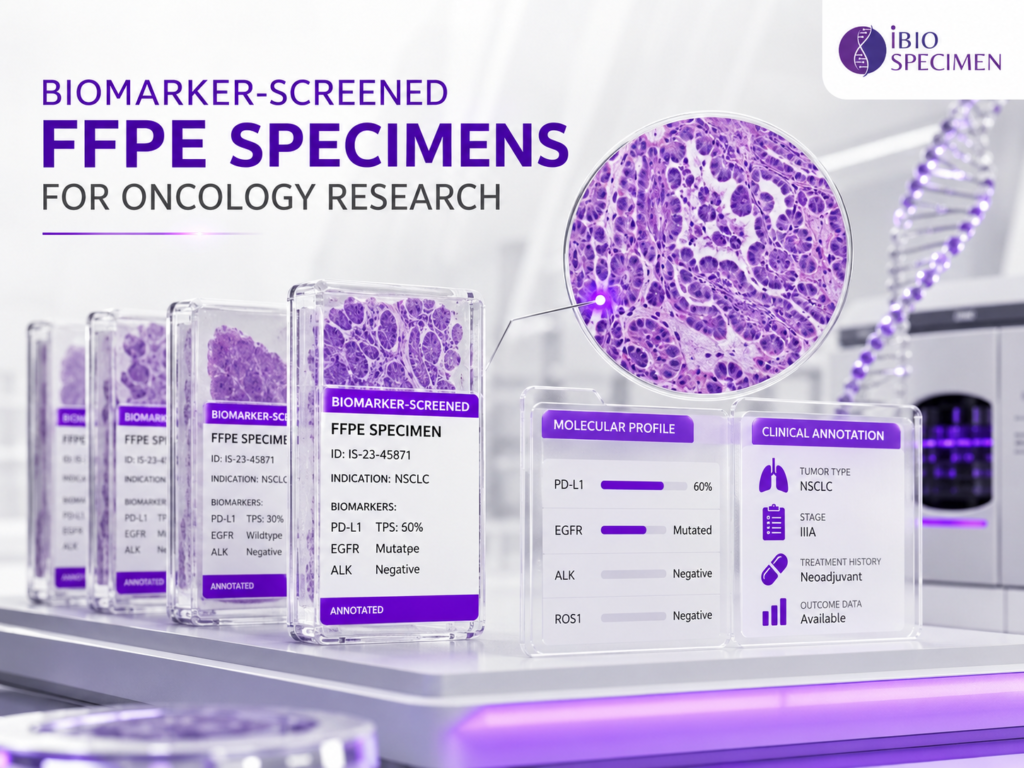
Modern oncology programs increasingly rely on AI Training Pathology Dataset to connect tissue phenotype with molecular biology, biomarker status and clinically meaningful research variables. In practice, the resource may combine H&E whole-slide images, digital pathology, expert pathology review, tissue morphology, depending on the study objective. The design should reflect whether the priority is broad discovery, disease-specific analysis, model pretraining, biomarker enrichment or independent validation. A useful starting point is to define the biological question first and then decide which images, specimens and metadata are required to answer it.
Cases can be organized by indication, histologic subtype, stage, grade, specimen type, collection period and other protocol-defined variables. A consistent data dictionary reduces downstream engineering work by defining units, permissible values, missing-data conventions and relationships between case-level and specimen-level fields. Structured identifiers are important because images, blocks, molecular results and clinical variables must remain linked to the correct donor and specimen without ambiguity. For molecularly focused studies, cases may be selected by mutation, copy-number alteration, expression profile, immunohistochemistry result or another pre-specified biomarker.
Pathologist Review, Annotation and Quality Control for AI Training Pathology Dataset
For pharmaceutical, biotechnology and AI teams, AI Training Pathology Dataset is most valuable when it is built as a study-ready resource rather than a loose collection of files or specimens. Quality is created through consistent linkage between H&E whole-slide images, digital pathology, expert pathology review and a structured case record. Each case should have a clear provenance trail showing how the diagnosis, specimen, digital asset and derived measurements relate to one another. Pathology and metadata review are particularly important before model training or downstream statistical analysis begins.
Conversely, tightly controlled cohorts can be assembled when the scientific question requires a narrow biomarker-defined or treatment-defined population. For image-centric programs, high-resolution whole-slide scans can be accompanied by slide-level labels, region annotations, tissue masks or cell-level measurements depending on the model objective. Where appropriate, pathology review can confirm diagnosis, tumor content, necrosis, tissue adequacy and the relationship between the specimen and the corresponding digital image. Cases can be organized by indication, histologic subtype, stage, grade, specimen type, collection period and other protocol-defined variables.
Using AI Training Pathology Dataset for Biomarker Discovery and Computational Oncology
AI Training Pathology Dataset can serve as a practical bridge between archived clinical material and modern oncology research when the data are curated around a clearly defined use case. AI Training Pathology Dataset can support research questions that are difficult to address with a single data modality. By combining H&E whole-slide images, digital pathology, expert pathology review, tissue morphology, investigators can study relationships that would otherwise remain hidden in separate data silos. The most valuable applications are those in which the cohort definition and analytical endpoint are specified before large-scale data generation begins.
For image-centric programs, high-resolution whole-slide scans can be accompanied by slide-level labels, region annotations, tissue masks or cell-level measurements depending on the model objective. Researchers can investigate associations between tissue morphology and genomic alterations, protein expression, gene expression or clinical outcomes. Pharmaceutical teams can use the resource for retrospective translational studies, exploratory biomarker work, cohort enrichment and hypothesis generation around drug response. When multiple modalities are linked at case level, the same cohort can support both image-based analysis and integrated computational biology workflows.
Custom AI Training Pathology Dataset Sourcing for Pharma, Biotech and AI Teams
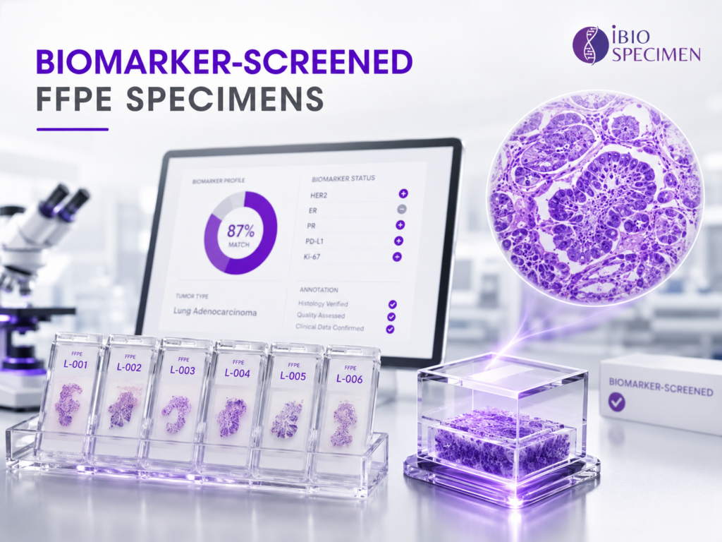
A high-value research dataset is useful only when the underlying cases are scientifically coherent, traceable and structured for the intended research question. Commercial development of AI Training Pathology Dataset should be approached as a controlled sourcing and data-engineering program rather than a one-time file transfer. The specification can define target indications, sample counts, biomarker groups, slide requirements, metadata fields and acceptance criteria before case identification starts. A pilot batch is often useful for checking the practical fit between the source material and the receiving team’s analytical pipeline.
Structured identifiers are important because images, blocks, molecular results and clinical variables must remain linked to the correct donor and specimen without ambiguity. Commercial projects may also require clear documentation of permitted research use, data handling expectations, de-identification approach and any limitations on redistribution. For multi-site programs, harmonized naming, metadata standards and quality thresholds are especially important because local laboratory practices may differ. A staged workflow—feasibility, pilot, QC review and scale-up—helps reduce the risk of building a large dataset that later proves inconsistent with the model or study requirements.
General Questions
Frequent Asked Questions!!
FFPE blocks for genomics are formalin-fixed, paraffin-embedded tissue samples used for DNA, RNA, and biomarker analysis. They are widely used in cancer genomics, molecular pathology, translational research, and retrospective studies.
FFPE tissue blocks are valuable because they preserve tissue architecture and molecular material for long-term storage. Researchers can use them for sequencing, mutation analysis, biomarker discovery, and validation studies.
Yes. DNA can be extracted from FFPE blocks using validated extraction kits and optimized laboratory protocols. DNA quality depends on fixation time, block age, tissue type, tumor content, and storage conditions.
Yes. RNA can be extracted from FFPE tissue, although it is often fragmented because of formalin fixation. Specialized FFPE RNA extraction methods can provide material suitable for targeted RNA sequencing, gene expression studies, and fusion analysis.
Yes. High-quality FFPE blocks are commonly used for next-generation sequencing, including targeted sequencing panels, whole-exome sequencing, RNA sequencing, and selected whole-genome applications.
Tumor content requirements depend on the study design and testing method. Many molecular and NGS studies require at least 20% tumor content, while some projects may require 30%, 50%, or higher tumor percentage. Pathologist review can be performed to confirm tumor content before shipment
FFPE blocks can support mutation testing, copy number analysis, gene fusion detection, microsatellite instability testing, tumor mutational burden analysis, methylation studies, and targeted DNA or RNA sequencing.
Yes. FFPE cancer tissue blocks are extensively used to study genomic alterations in lung, breast, colorectal, prostate, ovarian, pancreatic, liver, kidney, and other tumor types.
Researchers can purchase FFPE blocks from qualified biospecimen suppliers, biobanks, pathology laboratories, hospitals, and research networks that provide ethically sourced and clinically annotated human tissue samples.
Researchers should confirm diagnosis, tissue type, tumor percentage, necrosis percentage, fixation details, block age, specimen size, available clinical data, pathology review, consent status, and intended research-use permissions.
Yes. Clinically annotated FFPE blocks may include donor age, sex, diagnosis, grade, stage, TNM classification, treatment history, pathology report, mutation status, and clinical outcome data.